


MitoSpy 系列粒腺體活細胞染劑 Mitochondrion Living Cell Dye
MitoSpy™ 系列粒腺體活細胞染劑 Mitochondrion Living Cell Dye

MitoSpy™ 粒線體定位探針是可滲透細胞的螢光化學試劑,用於標記活細胞中的粒線體。
MitoSpy™ 根據其膜電位定位到粒線體,可用於指示細胞健康狀況以及定位,也可以被固定和透化以用於進一步的基於抗體的檢測。MitoSpy™ Orange CMTMRos 和 MitoSpy™ Red CMXRos 這兩種染劑適用於固定和透化的組織,亦可以與其他的抗體共染(co-stain)。
MitoSpy™ Orange 和 MitoSpy™ Red 含有一個氯甲基基團(chloromethyl group),以共價方式附著在細胞中的半胱胺酸(cysteines)上。這使它們在樣本固定(fixation)和透化(permeabilization)處理後更有可能保留在細胞中。然而,由於固定/透化過程會沖洗掉大量試劑,因此需要更高濃度的 MitoSpy™ Orange 和 MitoSpy™ Red 確保穩染色的穩定性。
在固定/透化後,大多數的 MitoSpy™ Green 和 MitoSpy™ NIR 將被沖洗掉。因此,在需固定/透化的樣本中不建議使用 MitoSpy™ Green 和 MitoSpy™ NIR 染劑。
BioLegend 昶安獨家代理
MitoSpy™ 細胞活染示意圖

| 
|
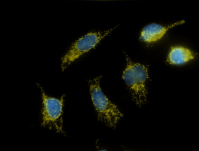
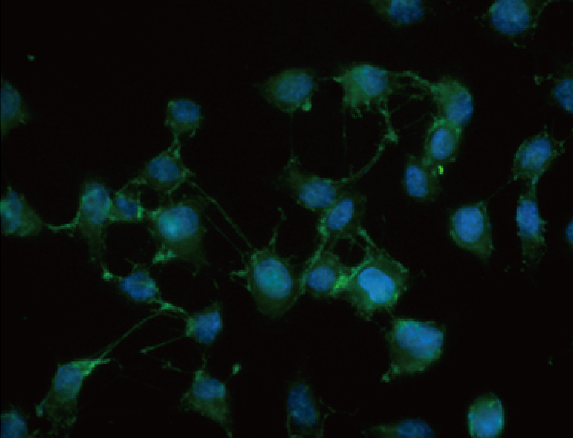
HeLa cells were stained with 0.25µM CytoPhase™ Violet dye (red) for 60 minutes at 37°C. Then 20 nM of MitoSpy™ NIR DiIC1(5) (green) was added for an additional 30 minutes at 37°C. The Z-stack images were captured on an LSM 880 with Airyscan using a 63x Oil objective. The files were Airyscan processed and a Maximum Intensity projection was created using Zen software; image courtesy of the Biophotonics Core Facility at the Salk Institute. | NIH3T3 cells were stained with 100 nM of MitoSpy™ Red CMXRos (red) for 20 minutes at 37°C, fixed with 1% PFA for ten minutes at room temperature, and permeabilized with 1X True Nuclear™ Perm Buffer for ten minutes at room temperature. Then the cells were stained with Flash Phalloidin™ NIR 647 (green) for 20 minutes at room temperature and counterstained with DAPI (blue). The image was captured with a 60x objective. |
BioLegend 昶安獨家代理
使用 MitoSpy™ 進行細胞活染
| Live staining (50 nM) |
PFA-fixed and permeabilized staining (500 nM) |
|
| MitoSpy™ Orange | 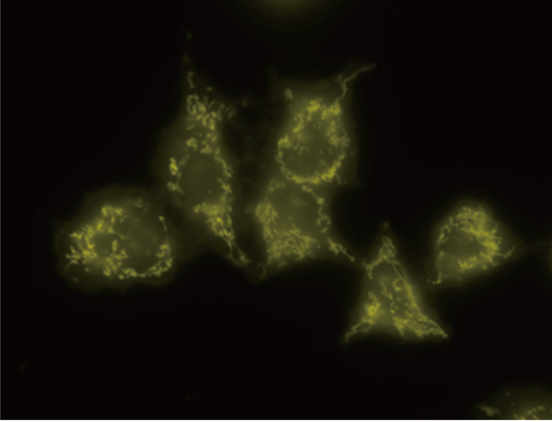 |
 |
| MitoSpy™ Green | 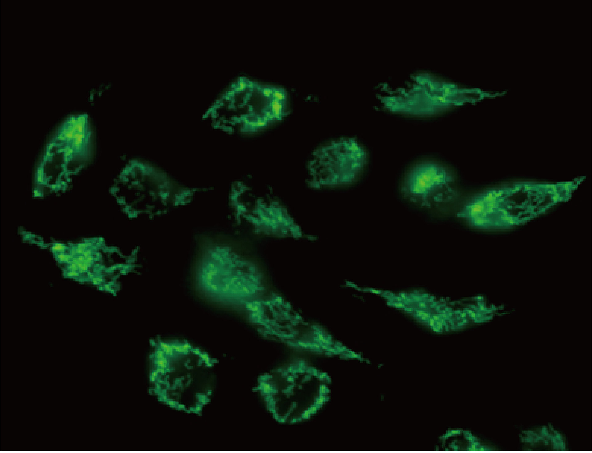 |
 |
HeLa cells that were stained live with either MitoSpy™ Orange (yellow) or Green (green). Cells that were fixed and permeabilized with 4% PFA and 0.1% Triton X-100 were also stained with DAPI (blue). Photos were taken with a 60x magnification. |
||
BioLegend 昶安獨家代理
MitoSpy™ 活染劑量建議
*請注意,所有MitoSpy™產品僅適用於活體樣本。以下建議反映了MitoSpy™是否能成功保留在「細胞處理」後。
| 細胞處理 | MitoSpy™ Orange 濃度建議 | MitoSpy™ Red 濃度建議 | MitoSpy™ Green 濃度建議 | MitoSpy™ NIR 濃度建議 |
|---|---|---|---|---|
| Live Cells | 50-250 nM | 50-250 nM | 50-250 nM | 1-50 nM |
| Fixed Cells | 50-250 nM | 50-250 nM | 50-250 nM | Not recommended. |
| Fixed and Permeabilized Cells | 250-500 nM | 250-500 nM | Not recommended. | Not recommended. |
BioLegend 昶安獨家代理
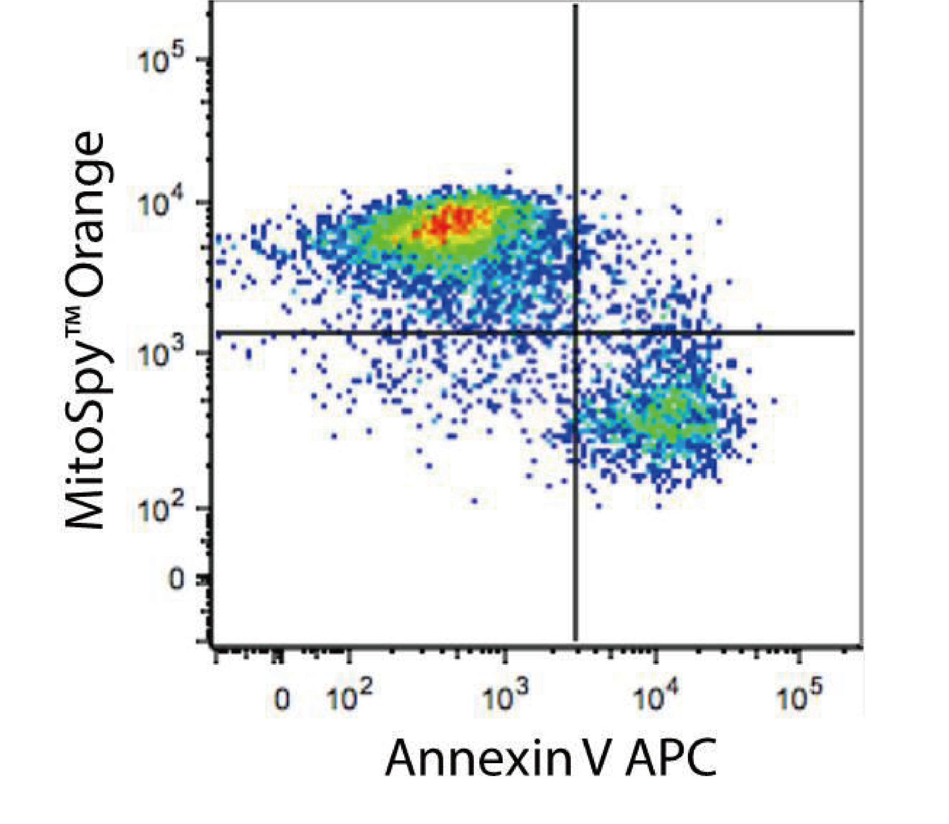
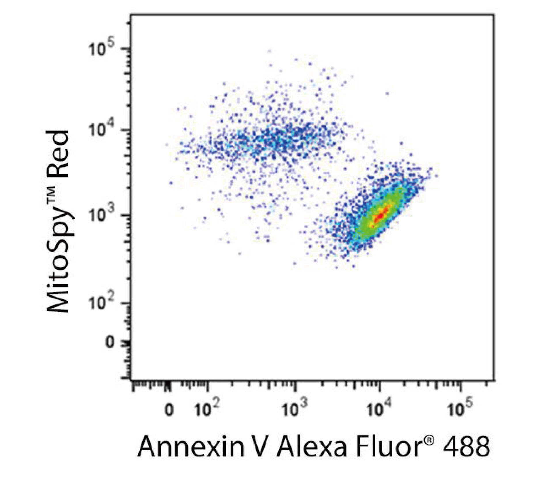
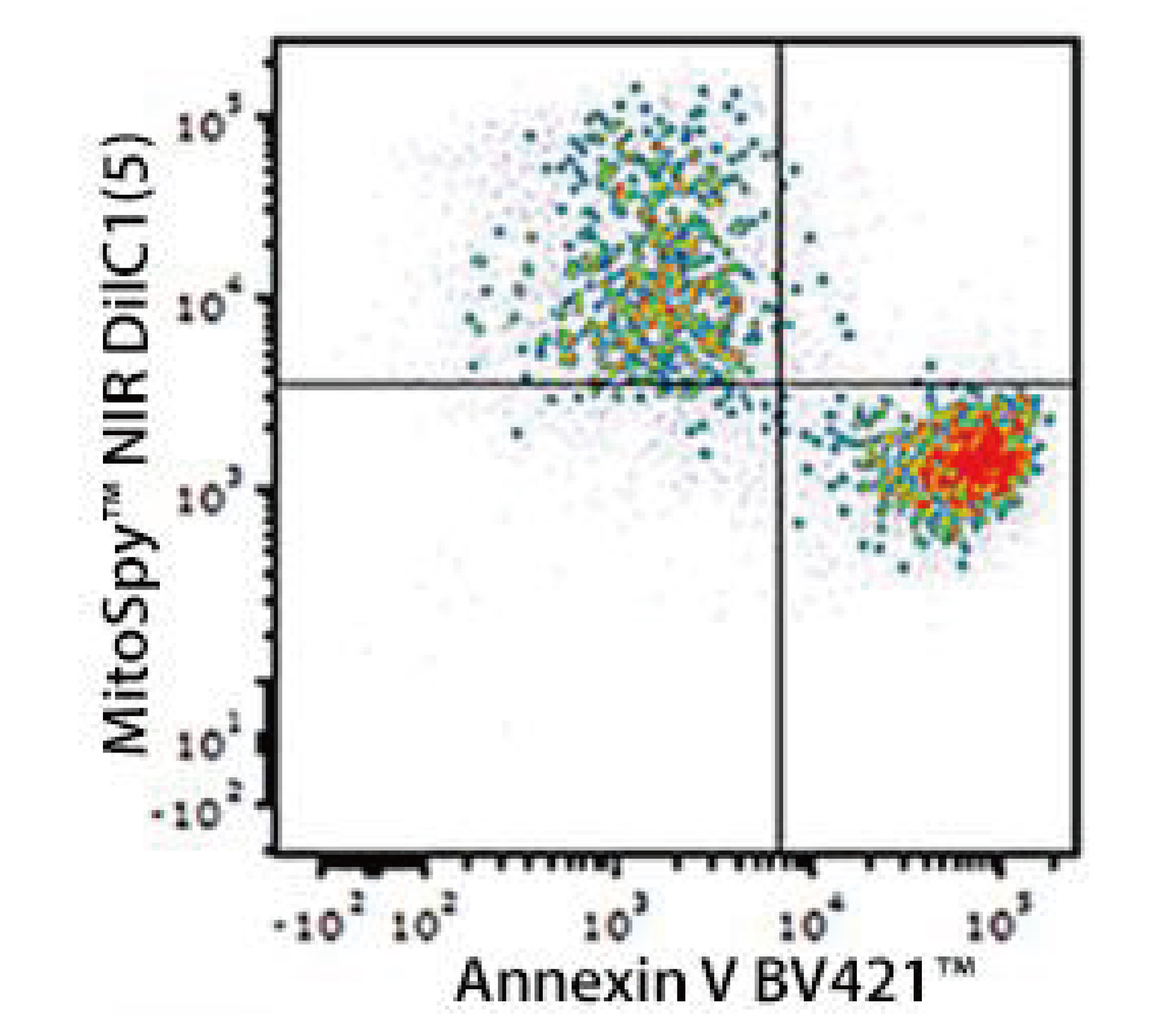
MitoSpy™ Flow Cytometry 染色示意圖
MitoSpy™ Orange、MitoSpy™ Red 和 MitoSpy™ NIR 也可用作細胞健康的指標。當細胞的粒線體正在進行主動的呼吸作用時,粒線體膜上會存在一個被稱為膜極化的電位差。如果細胞因為凋亡或細胞死亡而感到受損時,這個探針就不會強烈吸引到該細胞的粒線體。在下面的圖像中,對 MitoSpy™ Orange、MitoSpy™ Red 和 MitoSpy™ NIR 呈陽性的細胞是活躍且健康的,而對 Annexin V 呈陽性的事件處於凋亡的初期階段。在這種流式細胞儀應用中,MitoSpy™ Orange、MitoSpy™ Red 和 MitoSpy™ NIR 在分析前不應固定,因為固定會導致試劑的大量損失。低或負的 MitoSpy™ Orange、MitoSpy™ Red 或 MitoSpy™ NIR信號表明細胞具有凋亡表型。如果在固定過程中失去試劑,則信號強度的損失將混淆準確定性凋亡的能力。只有在成像應用中,MitoSpy™ Orange 和MitoSpy™ Red 才適用於亞細胞定位的固定。不建議使用 MitoSpy™ NIR 進行固定。
 | 
| 
|
Human T-cell leukemia cell line, Jurkat, was treated for 5 hours with LEAF™ purified anti-CD95 (clone EOS9.1), then stained with an impermeant nucleic acid stain, APC, Alexa Fluor® 488, or Brilliant Violet 421™ Annexin V, and either MitoSpy™ Orange, Red, or NIR as indicated. Nucleic acid stain positive events were excluded from analysis.
BioLegend 昶安獨家代理
MitoSpy™ 產品資訊
| 品名 | 激發光與散射光 | 共同光譜 | 反應物種 | 應用 | 貨號 |
| MitoSpy™ Green FM | 488 nm/520 nm | FITC, Alexa Fluor® 488 | Human, Mouse, Rat, All Species | ICC | 424805 |
| MitoSpy™ Orange CMTMRos | 551 nm/576 nm | PE, Alexa Fluor® 555, TRITC | Human, Mouse, Rat, All Species | ICC, FC | 424803 |
| MitoSpy™ Red CMXRos | 577 nm/598 nm | Texas Red®, Alexa Fluor® 594 | Human, Mouse, Rat, All Species | ICC, FC | 424801 |
| MitoSpy™ NIR DiIC1(5) | 638 nm/658 nm | Alexa Fluor® 647, APC | Human, Mouse, Rat, All Species | ICC (live imaging), FC | 424807 |
更多產品資訊,歡迎洽詢 BioLegend 台灣獨家代理 - 昶安科技。
點擊下列連接了解更多資訊: